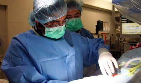
Structural heart conditions involve disease or defect in the heart's valves, walls and chambers. Our Structural Heart Program uses innovative, less-invasive procedures to treat these conditions and get you back to your life faster.
Our interventional cardiologists and heart surgeons work together with heart failure specialists and imaging experts to diagnose and treat structural heart conditions. We use minimally invasive techniques to repair heart valve problems, and this offers better outcomes, shorter hospital stays and faster recovery than open heart surgery.
The procedures do not require opening the chest, as a traditional surgery would require, and there is potential for discharge the following day.
Structural heart procedures
Our Structural Heart Program offers the following procedures.
Transcatheter aortic valve replacement (TAVR)
Transcatheter aortic valve replacement (TAVR) is a minimally invasive heart valve surgery that is used to treat aortic valve stenosis, which occurs when the aorta thickens and narrows, keeping the valve from fully opening and reducing blood flow to the body.
Mitral valve repair
The mitral valve separates the left chambers of the heart and lets oxygenated blood pass from the atrium to the ventricle. A leaky mitral valve causes the blood to flow backward from the left ventricle to the left atrium. If untreated, the condition can cause heart failure, irregular heart rhythm, stroke and sudden cardiac death.
Our Structural Heart Program offers a minimally invasive mitral valve repair procedure using the MitraClipTM device. This procedure is for patients with severe mitral valve leakage, known as mitral regurgitation, and is an alternative to open heart valve surgery. The procedure involves inserting a catheter into the femoral vein in the groin and advancing it to the heart using advanced imaging for guidance. The clip is threaded through the catheter and placed on the mitral valve leaflets, which brings them together, ensuring a better seal when the valve closes.
The procedure takes two to three hours. Patients usually can go home after an overnight stay in the hospital and can quickly return to daily activities.
Left atrial appendage (LAA) closure
The Left atrial appendage (LAA) closure procedure is a minimally invasive surgery that drastically reduces the chance of stroke for patients with nonvalvular atrial fibrillation (AFib).
The left atrial appendage — a small pouch connected to the upper left chamber of the heart — is where 90% of clots form in AFib patients. Using an LAAO closure device, such as the WATCHMANTM, our team of experts can block the left atrial appendage, reducing the risk of stroke. The device serves as a small umbrella, blocking the left atrial appendage.
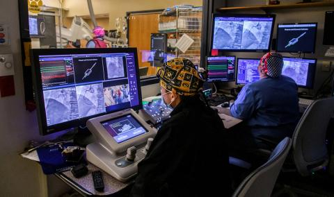
During an LAA closure procedure, a catheter is inserted in the femoral vein in the groin and threaded to the heart. The cardiologist, with the help of ultrasound imaging, guides the catheter tip to the left atrial appendage and deposits the device.
Patients who have this procedure are required to stay in the hospital overnight. After six weeks, patients have a follow-up visit to make sure the heart's lining has grown over the device, forming a solid barrier so clots can't escape. At that point, patients are prescribed the blood thinner Plavix for a few months and then can stop taking blood thinners altogether.
Related Conditions & Treatments
- Aortic Disease Care
- Atrial Fibrillation (AFib) and Arrhythmia
- Carotid Artery Disease
- Chest Pain
- Chronic Total Occlusion (CTO)
- Congenital Heart Disease
- Coronary Artery Disease
- Cardiothoracic Surgery
- ECMO Heart and Lung Life Support
- Heart Attack
- Congestive Heart Failure
- Heart Valve Disease
- Pediatric Cardiology
- Pediatric Vascular Anomalies
- Peripheral Artery Disease (PAD)
- Structural Heart Program
- Transcatheter Aortic Valve Replacement (TAVR)
- Vascular Surgery
- Women's Heart Health


